Fil:Surface projections of the organs of the trunk.png

Fuld opløsning(1.583 × 2.533 billedpunkter, filstørrelse: 3,33 MB, MIME-type: image/png)


Denne fil er fra Wikimedia Commons

Beskrivelse

Beskrivelse
English: Surface projections of the major organs of the trunk, using the vertebral column and rib cage as main reference points of superficial anatomy. The transpyloric plane and McBurney's point are among the marked locations.

To discuss image, please see Talk:Human body diagrams

Dato
Kilde

All included images are in public domain. See Template:Human body diagrams for individual details.

References

Spinal vertebrae levels:

  • Located at middle of frontal face of spinal vertebrae according to reference the image :File:Gray 111 - Vertebral column.png when pasted into image to superiorly fit the surface bulges of the clavicular bones and inferiorly fit the surface bulges of the anterior iliac spine.

Gross overview of organ locations:

  • Plate 260B - Regions and Planes of Adbomen. From: The Interactive Atlas of Human Anatomy Version 3.0 (CD-ROM). Authors: Frank H. Netter and Carlos A. G. Machado. Published by Saunders, October 1, 2002. ISBN-10: 1929007140. ISBN-13: 978-1929007141.
  • Fig 139 from The SPENCER SYSTEM. SEVENTEENTH EDITION
  • Plate 1034 from Gray's Anatomy

Locations of specific organs:
The following vertebral levels are generally given by the middle of the vertebral body.

  • Bålens ytanatomy (Superficial anatomy of the trunk). Anca Dragomir, Mats Hjortberg and Godfried M. Romans. Section for human anatomy at the Department of medical cell biology, Uppsala university, Sweden.
Key points from that compendium:
  • The oblique fissure of the right lung goes from the intercostal space 4 to 6
  • The horizontal fissure goes along the 4th rib (the original image from Gray has this line almost at the 5th rib, but this is not labeled in image, as other lung images seem to support the upper level)
  • The cardia of the stomach is at the level of the 10th rib
  • The fundus of the stomach is in the 5th intercostal space, slightly below the apex of the heart
  • Pylorus is usually at the L1 (the transpyloric plane)
  • The transpyloric plane is at the level of:
    • lumbar vertebra 1
    • the pancreatic body
    • the origins of the superior mesenteric artery from the aorta and portal vein
    • the left and right colic flexure
    • hilum of the kidney on the left
    • upper pole of the kidney on the right
    • duodenojejunal flexure
  • McBurney's point is located one third of a line from the anterior superior iliac spine and the navel
  • The upper rim of the liver is in the 5th intercostal space
  • The left lobe of the liver is to the left of the midline
  • The lower limit of the liver at the midline is approximately 3 cm below processus xiphoideus
  • The spleen is projected against the 9th and 10th rib on the backside
  • The left kidney is approximately at the vertebral level T12 to L3, and the right one slightly lower.
  • Atlas of Human Anatomy in Cross Section: Appendix: Topography of the Thorax and Abdomen. Ronald A. Bergman, Ph.D., Adel K. Afifi, M.D., Jean J. Jew, M.D., and Paul C. Reimann, B.S. [1]:
  • The average position of the highest point of the spleen is at the level of the lower third of the tenth thoracic vertebra.
  • The average position of the lowest point of the spleen is at the level of the middle third of the first lumbar vertebra.

Position of the navel

Urinary tract:

Forfatter Mikael Häggström

Licensering

Public domain Jeg, indehaveren af ophavsretten til dette værk, udgiver dette værk som offentlig ejendom. Dette gælder i hele verden.
I nogle lande er dette ikke juridisk muligt. I så fald:
Jeg giver enhver ret til at anvende dette værk til ethvert formål, uden nogen restriktioner, medmindre sådanne restriktioner er påkrævede ved lov.

Human body diagrams

Main article at: Human body diagrams

Template location:Template:Human body diagrams

How to derive an image

Derive directly from raster image with organs

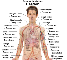
The raster (.png format) images below have most commonly used organs already included, and text and lines can be added in almost any graphics editor. This is the easiest method, but does not leave any room for customizing what organs are shown.

Adding text and lines:

Derive "from scratch"

By this method, body diagrams can be derived by pasting organs into one of the "plain" body images shown below. This method requires a graphics editor that can handle transparent images, in order to avoid white squares around the organs when pasting onto the body image. Pictures of organs are found on the project's main page. These were originally adapted to fit the male shadow/silhouette.

Organs:

More organs are found at: Human body diagrams/Organs

Derive by vector template

The Vector templates below can be used to derive images with, for example, Inkscape. This is the method with the greatest potential.

See Human body diagrams/Inkscape tutorial for a basic description in how to do this.

Examples of derived works

More examples

Licensing

Creative Commons CC-Zero Denne fil er udgivet under Creative Commons CC0 1.0 Universal Public Domain Dedication.
Personen, der har tilknyttet et værk til dette dokument, har dikteret værket som offentlig ejendom ved at give afkald alle hans eller hendes rettigheder til værket på verdensplan efter lov om ophavsret og alle relaterede og beslægtede rettigheder, i det omfang lovgivningen tillader det. Du kan kopiere, distribuere og forarbejde værket selv til kommercielle formål; alt sammen uden at spørge om godkendelse.

Captions

Tilføj en kort forklaring på en enkelt linje om hvad filen viser

Elementer som er med i denne fil

afbilder

image/png

Filhistorik

Klik på en dato/tid for at se filen som den så ud på det tidspunkt.

(Nyeste | Ældste) Se (10 nyere | ) (10 | 20 | 50 | 100 | 250 | 500)
Dato/tidMiniaturebilledeDimensionerBrugerKommentar
nuværende27. dec. 2019, 11:19Miniature af versionen fra 27. dec. 2019, 11:191.583 × 2.533 (3,33 MB)Mikael Häggström+Costal margin
11. nov. 2010, 12:38Miniature af versionen fra 11. nov. 2010, 12:381.050 × 1.680 (2,07 MB)Mikael HäggströmAdapted to recently added overview images. Distinguished different ways to designate vertebrae levels.
7. nov. 2010, 12:04Miniature af versionen fra 7. nov. 2010, 12:04936 × 1.325 (1,77 MB)Mikael Häggströmupdate from svg
7. nov. 2010, 11:46Miniature af versionen fra 7. nov. 2010, 11:46936 × 1.325 (1,77 MB)Mikael Häggströmupdate from svg
24. okt. 2010, 06:51Miniature af versionen fra 24. okt. 2010, 06:51936 × 1.325 (1,61 MB)Mikael HäggströmSmoother edges
10. okt. 2010, 07:18Miniature af versionen fra 10. okt. 2010, 07:18936 × 1.325 (1,61 MB)Mikael HäggströmMinor kidney adjustment. More realistic hip bone
6. okt. 2010, 06:47Miniature af versionen fra 6. okt. 2010, 06:47936 × 1.325 (1,73 MB)Mikael HäggströmDistinguished stomach and spleen. Removed painted arteries out of scope.
4. okt. 2010, 20:40Miniature af versionen fra 4. okt. 2010, 20:40936 × 1.325 (1,74 MB)Mikael HäggströmLowered spleen
3. okt. 2010, 17:21Miniature af versionen fra 3. okt. 2010, 17:21936 × 1.325 (1,74 MB)Mikael HäggströmDecreased some opacity. Aligned tail of pancreas with spleen. Adjusted fissure marking width.
2. okt. 2010, 20:20Miniature af versionen fra 2. okt. 2010, 20:20936 × 1.325 (1,72 MB)Mikael Häggström+liver label
(Nyeste | Ældste) Se (10 nyere | ) (10 | 20 | 50 | 100 | 250 | 500)

Den følgende side bruger denne fil:

Global filanvendelse

Følgende andre wikier anvender denne fil:

Vis flere globale anvendelser af denne fil.

Metadata